

Aortic Pulse Wave Velocity (PWVao)
The most frequently used parameter in the medical scientific literature for describing arterial stiffness is pulse wave velocity (PWV). PWV is mainly related to aortic wall characteristic. Basically, the more rigid the aortic wall, the faster the aortic PWV.
Aortic Pulse Wave Velocity (PWVao) is a key predictor of coronary heart disease and stroke in healthy subjects without symptoms. It is an important marker of cardiovascular risk and is increasingly used in medical practice nowadays. Aortic PWV is also associated with early signs of coronary atherosclerosis, independent of other risk factors like blood pressure.
What is Pulse Wave Velocity?
Aortic PWV is the speed at which the pulse pressure travels through the aorta. It is used in clinics to assess arterial stiffness and can be measured non-invasively with an Arteriograph. In fact, measuring PWV provides strong evidence of large artery stiffness. Higher PWVao can predict cardiovascular death and overall mortality in the general population, specifically in individuals suffering from end-stage kidney disease, hypertension, and diabetes
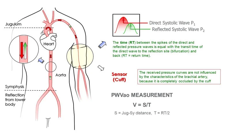
Arteriograph is equipped with an inflatable cuff placed on the patient’s upper arm and inflated 45 mmHg above the individual’s Systolic Blood Pressure (SBP). Pressure variations are detected by a pressure sensor and signal is transferred to a computer. PWV acquirement is based on the generation of two systolic peaks: the first peak (P1) results from the systolic volume ejection in the aorta, whereas the second and lower peak P2 is given by wave pressure reflection from peripheral arteries. Return time is the difference between the first peak (P1) and the reflected systolic peak (P2). PWV is calculated as shown:
PWV = S (Jug-Sy)/(RT/2)
Distance (S) is measured from jugular (Jug) to symphysis (Sy), RT: return time
What is Normal Pulse Wave Velocity?
As age increases, pulse wave velocity (PWV) often rises. However, studies indicate that aortic pulse wave velocity (PWVao) is typically considered normal when it remains below 10 m/s. It is worth noting that most devices currently available, except for the Arteriograph, measure other forms of PWV, which have faced criticism in various scientific studies. Among these methods, aortic PWV is widely recognized as the most reliable and is regarded as the gold standard for assessing arterial stiffness.